The Best Microscopic Shots of 2025 Will Make You Rethink Reality
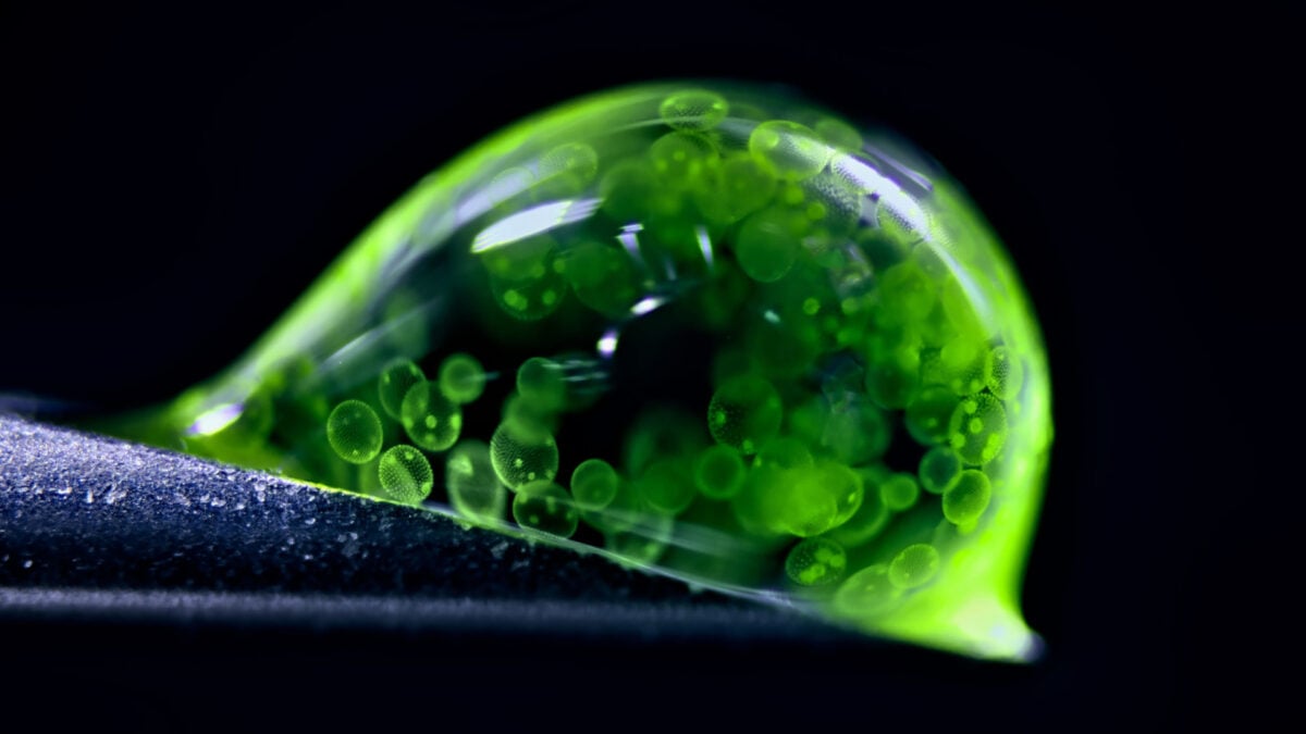
What you see here isn’t slime, nor jelly. It’s a photograph of an extremely zoomed-in drop of water housing tiny spheres of colonial algae, which Jan Rosenboom, a chemical engineer from Germany, captured using reflected light microscopy.
Rosenboom’s image was the runner-up of Nikon’s annual Photomicrography Competition, hosted by the company to honor the work of microscopy in science. Every year, professional scientists and casual enthusiasts rise to the occasion, sharing mind-blowing snapshots of familiar objects seen from entirely new angles. (Our own Ed Cara was a judge for the competition in 2023.)
This year’s winners range from close-ups of human cellular networks and the hidden worlds inside unassuming mushrooms to microscopic creatures you probably didn’t even know existed. You can check out the full list here, but we’ve picked some of our favorites for your viewing pleasure.
Red fungal pigments
Fungi may be some of the strangest life forms on Earth, but photomicrography showcases how pretty they can get. Wim van Egmond from the Micropolitan Museum in the Netherlands placed ninth in the competition for his stunning close-up of red, diffused pigments of the fungus Talaromyces purpureogenus, a distant relative of the penicillin-producing Penicillium.
Fluorescently marked mouse colon
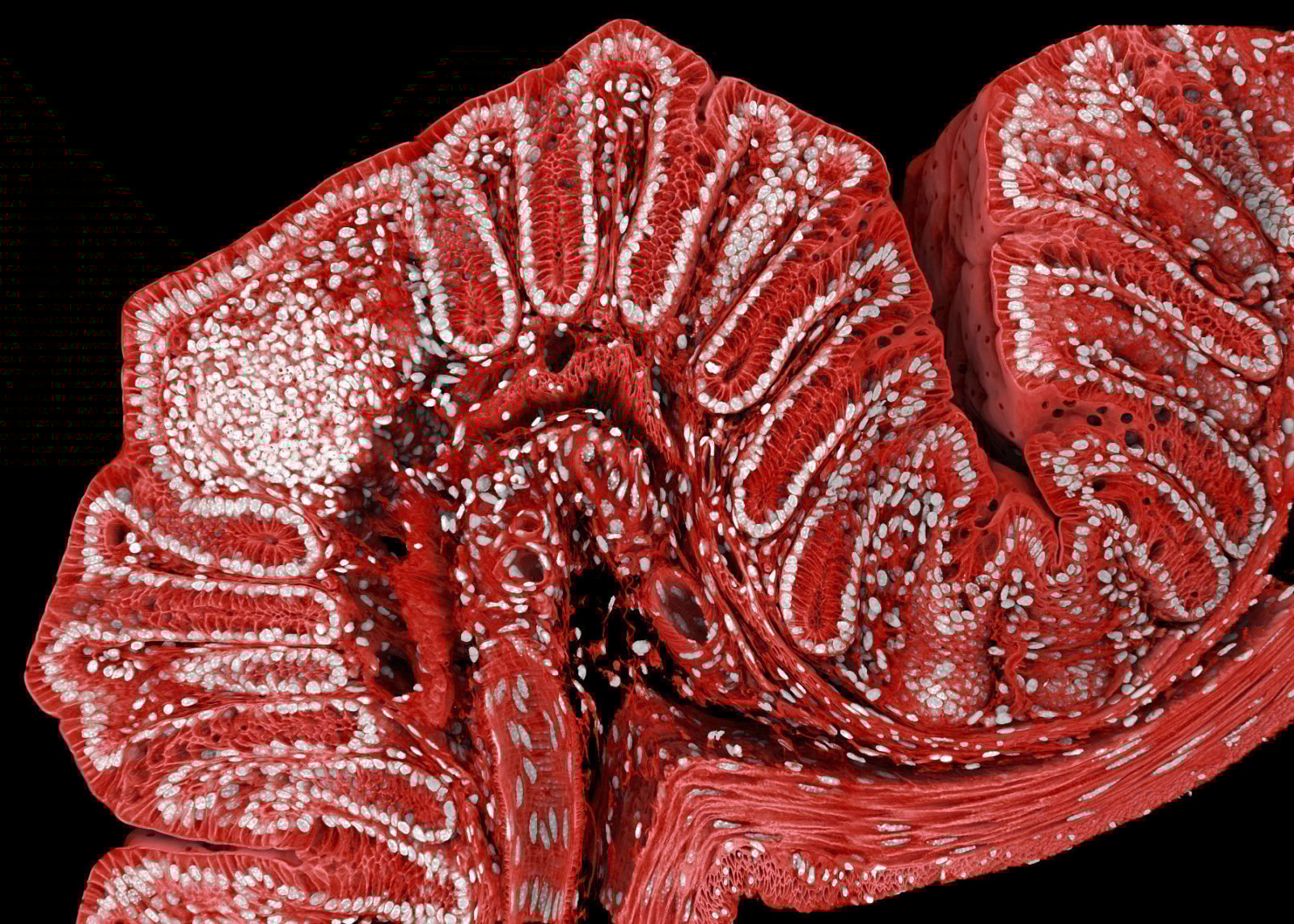
Mice play a big role in scientific research. This image of the mouse colon taken by researchers at the Friedrich Miescher Institute for Biomedical Research in Switzerland used confocal microscopy, a popular technique in the biomedical sciences for studying cells marked with fluorescent probes.
Heart cells at work
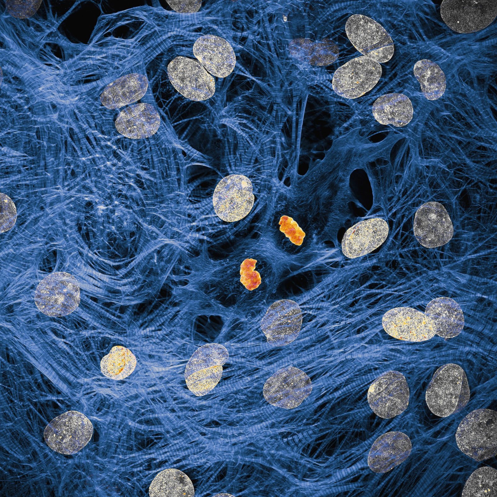
You don’t have to be an expert to know that the cellular networks inside our bodies work diligently to ensure everything functions well. James Hayes of Vanderbilt University captured heart muscle cells with chromosomes condensing after cell division.
The galaxy inside your head
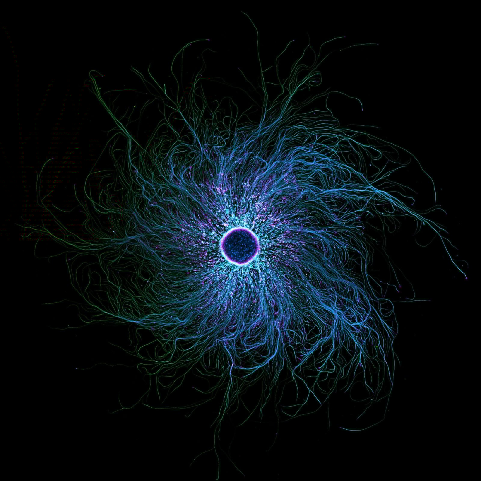
Sometimes, pictures of the microscopic world betray first impressions. Although this image strongly resembles a frenzied black hole, the subject of the photograph, taken by Stella Whittaker of the NIH, is iPSC-derived sensory neurons, labelled to show two proteins, tubulin and actin. Whittaker used a combination of microscopy techniques for this shocking image.
Parasite larvae
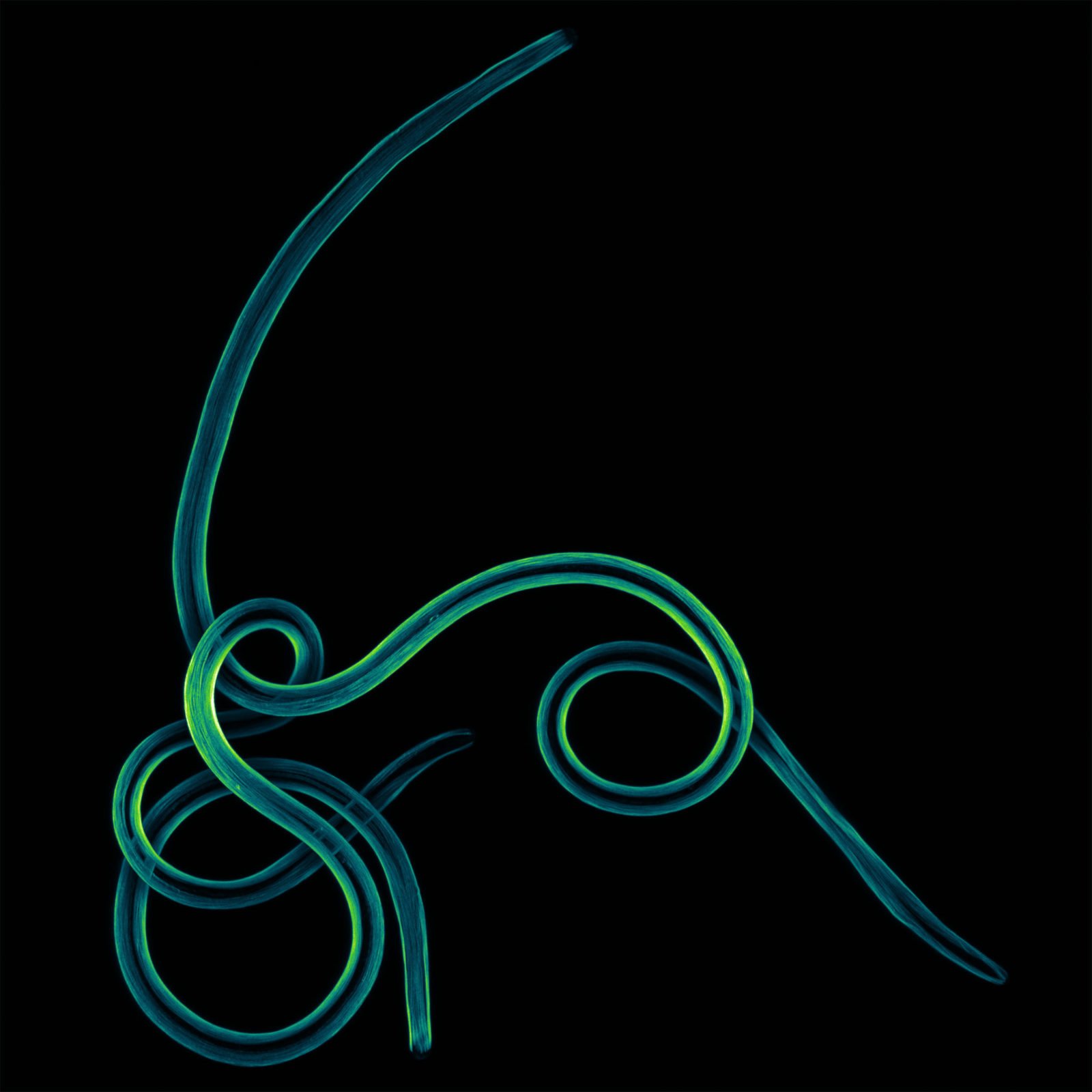
Filariasis refers to a parasitic infection caused by the pictured specimen, a filarial nematode parasite. The tropical disease causes painful rashes, malfunctioning cells, and even blindness. Up close, though, it sure looks far from threatening.
Pollen caught in a web
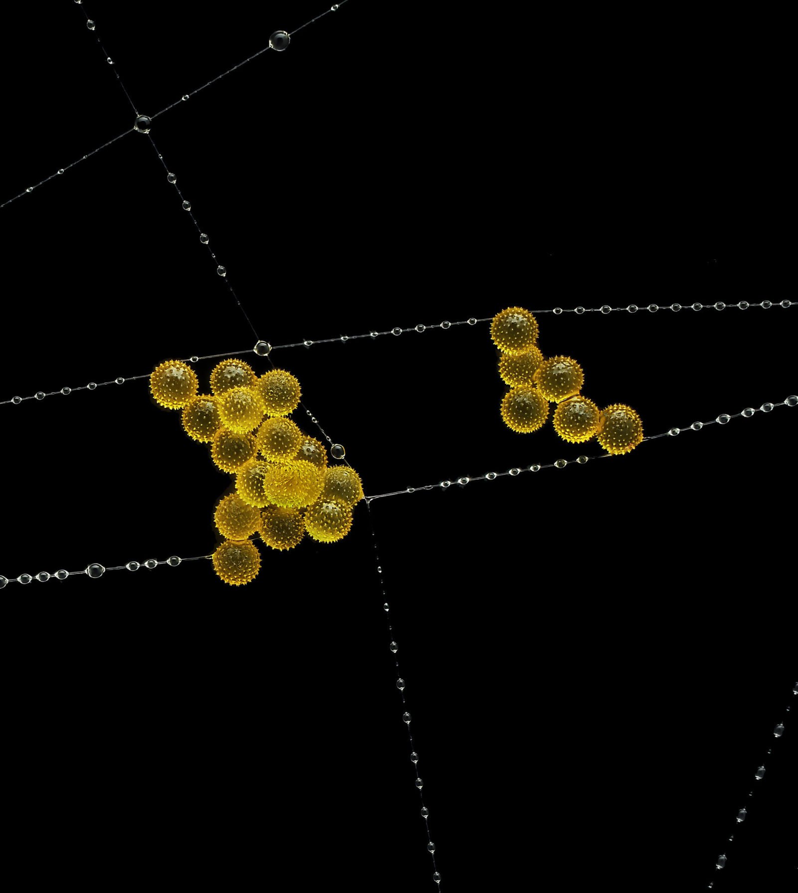
Nature is full of delicate yet surprisingly sturdy structures—as represented by this striking image of pollen spores literally hanging by a thread on a garden spider web. John-Oliver Dum from Germany’s Medienbunker Produktion won third place for his photograph, a combination of multiple shots stacked atop each other.
A three-way feast?
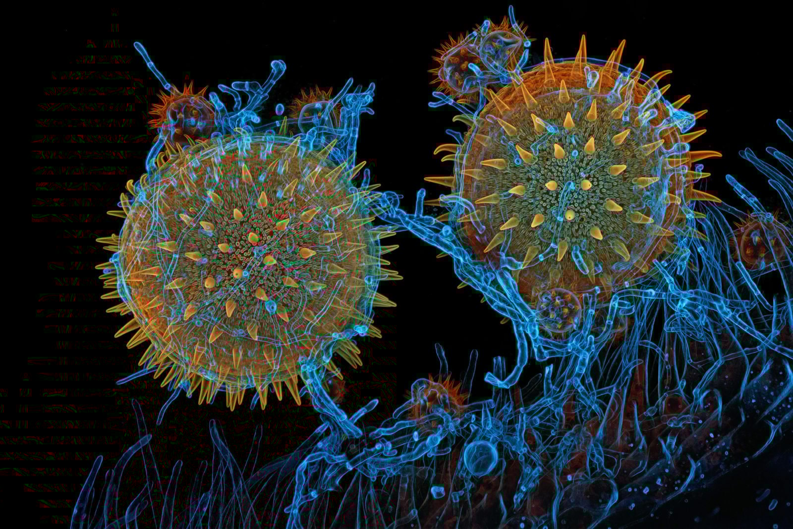
Then again, nature can also get really brutal, and extreme close-ups make that even clearer. Igor Robert Siwanowicz of the Howard Hughes Medical Institute caught marrow pollen “germinating on stigma while being parasitized by a filamentous fungus,” illustrating the wild interdependent relationships of the microscopic world.
Rice weevil on a grain of rice
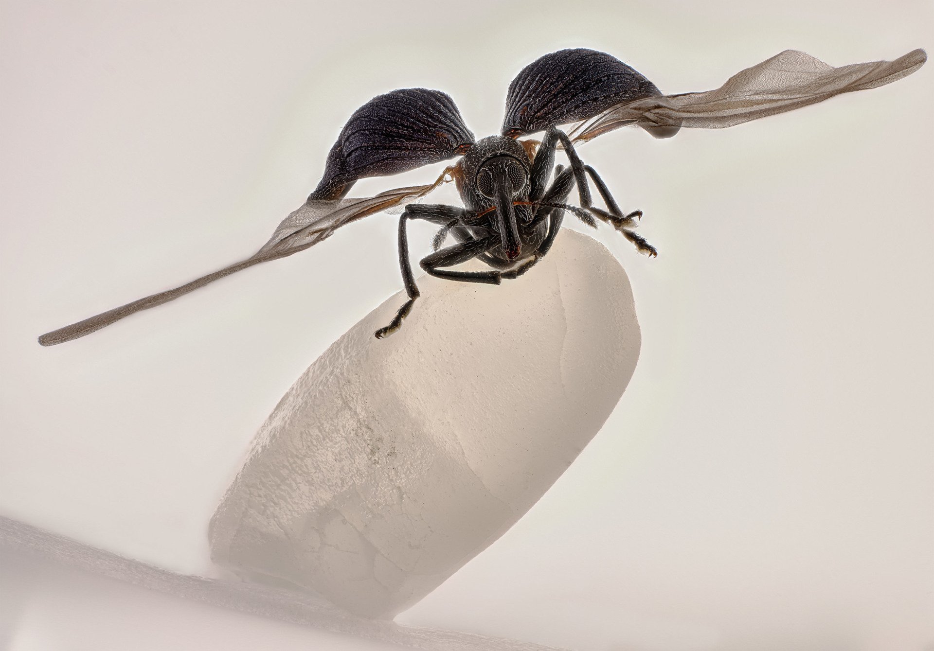
Last but certainly not least, the overall winner of the competition caught a rare moment of a rice weevil spreading its wings while perched on one grain of rice. The photograph, this year’s overall winner, is actually a combination of over 100 images stacked, cleaned, and post-processed to maximize clarity and impact.
Zhang You, an entomologist from China, said in his winning statement that the image “is a product of the years…spent focused on ecological and insect science photography, as well as teaching others about entomology.”
First Appeared on
Source link

