Sci-fi style 4D ultrasound breakthrough reveals full organ blood flow
A new ultrasound-based imaging tech has been developed to map the organ blood flow in four dimensions (3D + time) — a level of detail previously unattainable.
This new medical imaging tech could provide deeper insights into the circulatory system as well as enhance the diagnosis and treatment of blood circulation-related diseases.
The development comes from a team of Inserm researchers at the Physics for Medicine Institute (Inserm/ESPCI Paris-PSL/CNRS) in France.
For the first time, the new imaging technique mapped the 4D blood flow of entire organs in animal models.
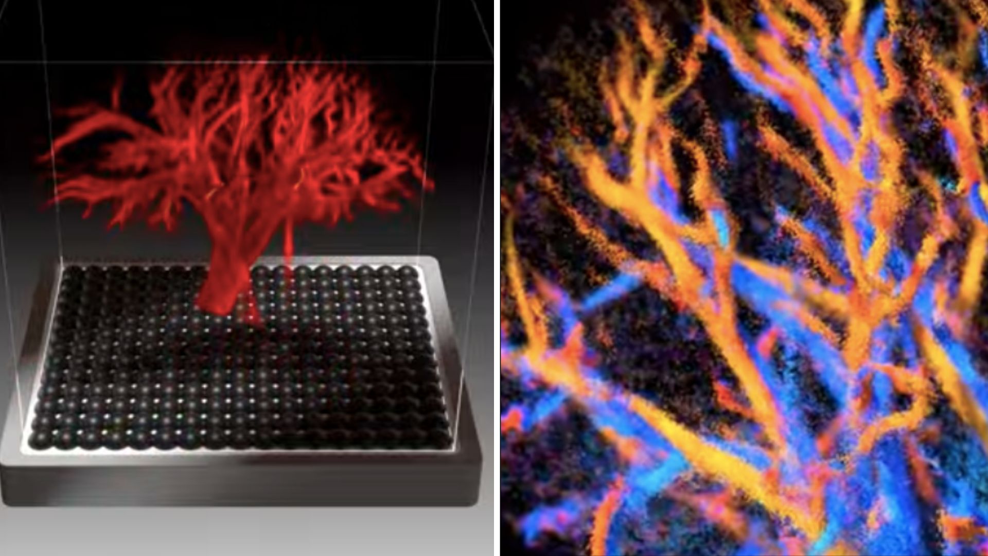
“The originality of these results lies in the fact that these images allow us to visualise the vessels of an entire organ at very small scales (less than 100 micrometres) – this 4D image resolution is unprecedented, as is the ability to observe an entire large organ and its flow dynamics,” explained Clément Papadacci, Inserm researcher and last author of the study.
The team released an illustration-based video of this new imaging technique on October 28.
Unprecedented view inside organs
The human circulatory system involves veins, arteries, vessels, and the lymphatic system.
It depends on a complex network of blood vessels to transport oxygen, nutrients, and waste. Crucial to this is blood microcirculation, which flows through tiny vessels that feed cells and remove metabolic waste.
Disruptions in this microcirculation, whether in structure or function, can lead to serious conditions like heart failure, kidney failure, and chronic diseases.
A key medical challenge has been the lack of a tool. No existing method could visualize the entire circulatory network across a whole organ, from large arteries to the smallest arterioles.
The team’s new non-invasive ultrasound probe is the first to solve this problem.
With an unprecedented image resolution, this technology led to mapping of the vascularization to precisely quantify the blood flow dynamics of the heart, kidney, and liver in animal models comparable to human size.
This non-invasive tool can distinguish microcirculation in the finest vessels under 100 micrometers.
In the liver, the technology showed its power. It differentiated the organ’s three distinct blood networks — arterial, venous, and portal. The tech analyzed their unique hemodynamic signatures (blood flow characteristics).
Future clinical applications
This non-invasive technology holds great promise for patients. The researchers aim for easy adoption in clinics.
“The probe can be connected to small portable equipment, which would allow it to be integrated into medical practice,” noted Papadacci.
The device’s ability to observe an entire large organ and its flow dynamics with such precision is expected to be a game-changer.
The next step involves testing this technology in human clinical trials.
These developments are being expedited with the support of the Technological Research Accelerator for Biomedical Ultrasound, an initiative by Inserm integrated into the Physics for Medicine Institute.
Papadacci believes the tool will be widely used.
“Used in clinical settings, this new technology could become a major tool for better understanding vascular dynamics as a whole, from the largest vessels to the pre-capillary arterioles,” said Papadacci.
The technology holds potential to improve the diagnosis of microcirculation disorders. It will aid in monitoring treatments for small vessel diseases, which are difficult to diagnose and usually identified only after ruling out other pathologies.
The study was published in the journal Nature Communications.
First Appeared on
Source link

