How Slow Waves During Sleep Take Over to Clear Metabolic Trash
Summary: We’ve long known that sleep “washes” the brain, but we’ve never been able to see it happen in real-time without invasive dyes—until now. Researchers have developed an ultrafast MRI technique that tracks the movement of brain fluids non-invasively.
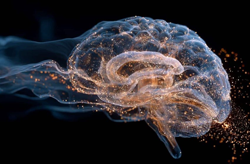
The study shows that during sleep, the brain’s “operating logic” actually reverses. Instead of neurons controlling blood flow, slow vasomotor waves (vascular pulsations) begin to drive both fluid movement and electrical activity, specifically in the sensory cortex, to flush out metabolic waste.
Key Facts
- Contrast-Free Tracking: The new “ultrafast MRI” method tracks water molecules in cerebrospinal fluid directly, requiring only a five-minute scan with no injected chemicals.
- The Pulse Shift: During sleep, respiratory and vasomotor pulsations (the “cleaning” waves) speed up, while cardiac pulsations (heartbeat waves) slow down, allowing for more efficient “filtering” of brain tissue.
- The Reverse Control: In an awake brain, neurons tell blood where to go. During sleep, the vascular waves take the lead, influencing the neurons and pushing fluid through the posterior brain regions to clear waste.
- Wearable Future: Beyond MRI, the team developed wearable sensors that can track these cleansing rhythms in a standard bed, opening the door for routine monitoring of the aging brain.
Source: University of Oulu
Sleep helps the brain to cleanse itself – and now this process can be measured in humans entirely non-invasively.
Researchers at the University of Oulu have developed a method that allows the increased movement of brain fluids during sleep to be tracked quickly and safely, without the need for injected contrast agents.
The brain’s cleansing mechanism is driven by pulsations, natural bodily rhythms that move blood and cerebrospinal fluid through the brain. These pulsations fall into three main categories: cardiovascular pulsations generated by the heartbeat in arteries, respiratory pulsations affecting veins and cerebrospinal fluid spaces, and slow vasomotor waves in the walls of blood vessels. Previous research has shown that both these pulsations and the brain’s waste clearance are enhanced during sleep.
These pulsations drive the flow of fluids through brain tissue, helping to remove metabolic waste. When this fluid circulation weakens, waste products may begin to accumulate in the brain. The phenomenon has been linked to memory disorders, among other conditions, but has been difficult to measure directly in humans.
Fluid flow accelerates during sleep
An ultrafast magnetic resonance imaging method developed by the University of Oulu’s functional neuroimaging research group (OFNI) now makes it possible to measure brain fluid circulation directly by tracking the movement of water molecules in cerebrospinal fluid. The scan takes only about five minutes and does not require contrast agents.
The researchers found that the behaviour of brain pulsations changes markedly during sleep. The propagation of respiratory and vasomotor pulsations—both of which promote brain-cleansing fluid circulation—speeds up, while cardiac pulsations slow down. This shift is thought to reflect more efficient water filtration in brain tissue, alongside a slowing of arterial pulse waves as blood vessels dilate and blood pressure decreases during sleep.
Brain control dynamics partly reverse during sleep
The studies also revealed a shift in the brain’s fundamental operating logic during sleep. When awake, electrical activity in neurons modulates blood flow and fluid movement: neural activation comes first, followed by increased blood flow. During sleep, however, this relationship is no longer strictly one-directional.
“During sleep, vasomotor waves in particular, slow pulsations below 0.1 hertz, begin to locally influence not only fluid movement but also the brain’s electrical activity,” explains Professor Vesa Kiviniemi, who led the research.
This effect is especially pronounced in posterior brain regions, such as the sensory cortex. These same areas also show a marked increase in fluid flow through brain tissue, pointing to enhanced clearance.
New possibilities for monitoring the ageing brain
The findings are based on two recently published studies, one in Advanced Science and the other in The Proceedings of the National Academy of Sciences (PNAS). Both studies involved measurements in healthy volunteers.
According to the researchers, the results provide a more detailed understanding of how and where sleep enhances the brain’s cleaning processes.
It is already known that brain fluid circulation declines with age. “New measurement methods open up possibilities to monitor—and in the future potentially treat—age-related changes in brain fluid dynamics,” says Kiviniemi.
The research group has also developed wearable technology that can track brain electrical activity and blood flow during sleep without the need for MRI. The results correspond well with MRI measurements, suggesting that brain cleansing could in future be monitored more easily in clinical settings.
The team is now working on ways to enhance the fluid circulation and pulsation mechanisms that weaken with age, with the aim of slowing down the effects of ageing on the brain.
Key Questions Answered:
A: When you’re awake, your brain’s “electrical grid” is too busy processing information. The study shows that sleep triggers a specific “pulse shift”: your blood vessels dilate and slow down, which creates the physical space and pressure needed for cerebrospinal fluid to rush in and “scrub” the tissue.
A: Exactly. If those slow vasomotor waves don’t get a chance to take over and drive fluid flow, metabolic waste products stay trapped in your brain tissue. This accumulation is linked to long-term memory disorders and that immediate feeling of cognitive “heaviness.”
A: The Oulu team created technology that monitors electrical activity and blood flow simultaneously. Because they now know exactly how these two signals synchronize during the “cleaning” phase, they can use external sensors to confirm if your brain is successfully hitting its “wash cycle” without needing a giant MRI machine.
Editorial Notes:
- This article was edited by a Neuroscience News editor.
- Journal paper reviewed in full.
- Additional context added by our staff.
About this sleep and neuroscience research news
Author: Meri Rova
Source: University of Oulu
Contact: Meri Rova – University of Oulu
Image: The image is credited to Neuroscience News
Original Research: Open access.
“Sleep alters neurovascular and hydrodynamic coupling in the human brain” by Tommi Väyrynen, Johanna Tuunanen, Heta Helakari, Ahmed Elabasy, Vesa Korhonen, Niko Huotari, Johanna Piispala, Mika Kallio, Maiken Nedergaard, and Vesa Kiviniemi. PNAS
DOI:10.1073/pnas.2510731123
Open access.
“Sleep Alters the Velocity of Physiological Brain Pulsations in Humans” by Ahmed Elabasy, Heta Helakari, Tommi Väyrynen, Zalán Rajna, Niko Huotari, Lauri Raitamaa, Ville Isokoski, Matti Järvelä, Mika Kaakinen, Johanna Piispala, Mika Kallio, Vesa Korhonen, Tapio Seppänen, Vesa Kiviniemi. Advances Science
DOI:10.1002/advs.202503745
Abstract
Sleep alters neurovascular and hydrodynamic coupling in the human brain
Sleep is essential for maintaining brain tissue homeostasis, which is facilitated by enhanced cerebrospinal fluid (CSF) solute transport. Infraslow (<0.1 Hz) vasomotion, CSF flow, and electrophysiological potential all increase during sleep, but their contributions as potential drivers of CSF flow in human brain remain unknown.
To investigate this, we measured these signals in healthy volunteers across sleep–wake states using functional MRI blood oxygen level-dependent (BOLD), electroencephalography, and functional near-infrared spectroscopy.
We then studied the directed coupling patterns between the three signals, using phase transfer entropy. In the awake state, electrophysiological potential and water concentration changes both predicted hemodynamic BOLD changes across whole brain, reflecting classical functional hyperemia.
During sleep, these interactions changed such that the net directionality was lost and the interactions became more bidirectional.
Our results show that in addition to neural changes during sleep, nonneural processes such as vasomotor-driven hydrodynamic waves start to gain more impact on human brain activity.
Abstract
Sleep Alters the Velocity of Physiological Brain Pulsations in Humans
Introduction: The clearance of brain metabolites increases during sleep, in association with increased spectral power of the three main cerebrospinal fluid (CSF) flow drivers: cardiovascular, respiratory, and vasomotor brain pulsations.
However, little is known about how the increased power of these pulsations affects the velocity and direction of fluid flow in the sleeping brain.
Objectives: To address this knowledge gap, we mapped the CSF oscillatory flow velocity in relation to the changing physiological pulsations in the brains of 22 healthy volunteers during sleep and waking.
Methods: We used the ultrafast magnetic resonance imaging sequence known as magnetic resonance encephalography (MREG) for tracing the pulsatile movement of water molecules inside the cranium. First, we conducted a phantom validation study with optical flow analysis to confirm that MREG accurately tracks pulsatile water molecule flow in a porous tissue medium.
Next, we obtained MREG recordings for mapping the three physiological pulsations without aliasing in the human brain across the awake and sleep states; we thereby quantified the brain-wide 3D velocity vectors (i.e., the velocity vs and 3D direction ) of each pulsation band, using comprehensive dense optical flow analysis during EEG-verified sleep in comparison to the awake state.
Finally, we assessed relationships among the spectral power of the physiological pulsations, their 3D velocity , and slow-delta EEG power, which is known to depict the increased interstitial volume during sleep.
Results: In our phantom study, dense optical flow analysis reliably detected water flow in tissue driven by external pulsations. In healthy volunteers, sleep increased flow velocities () of the pulsations by more than 20% in concert with elevations in respiratory pulsations and vasomotor waves, while the velocity of cardiovascular pulsations (vs) declined by the same percentage.
There was a significant anticorrelation between cardiac mean spectral power and slow delta EEG mean power, and a significant correlation between vasomotor mean spectral power and slow delta EEG mean power over the whole brain.
Conclusions: Phantom studies validated the optic flow analysis of fast MREG recordings. Sleep altered the 3D velocity dynamics of all neurofluidic brain pulsations in a manner consistent with increased interstitial space and greater fluid exchange, thus supporting the glymphatic model wherein physiological pulsations drive bulk flow during sleep.
First Appeared on
Source link

