Scientists Finally Reconstruct Little Foot’s Face From a 3.67-Million-Year-Old Crushed Skull
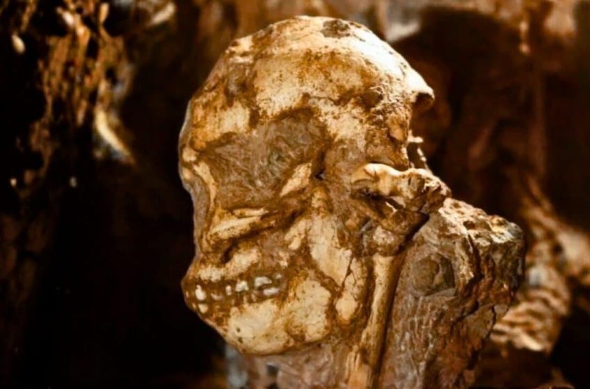
Technicians digitized a crushed fossil skull with enough precision to track each displaced fragment. The specimen was Little Foot, dated to 3.67 million years, and the scan was designed to solve a stubborn problem: the face had been warped so badly that physical reconstruction could not safely correct it. Researchers needed a way to rebuild the anatomy without forcing bone back into place.
The skull is catalogued as StW 573, excavated from the Sterkfontein Caves inside South Africa’s Cradle of Humankind. Little Foot is widely described as an exceptionally complete early hominin skeleton, yet the facial region stayed frustratingly unreadable because geological pressure bent and shifted key structures. That distortion kept the fossil from being compared cleanly with other early Australopithecus faces.
One widely read report on Earth.com followed the reconstruction step by step, starting with the basic obstacle: the facial bones existed, but not in their original alignment. The story’s core detail was not artistry, but measurement, because only a stable digital face could be treated as analyzable data rather than a fragile object.
A Fossil Landscape Built for Both Research and Visitors
Little Foot emerged from a cave system that is still an active research site, tied closely to the University of the Witwatersrand. The fossil’s location matters because Sterkfontein sits near Johannesburg, within a concentrated corridor of hominin discoveries that has shaped how scientists frame early human ancestry in southern Africa.
That same corridor also supports public interpretation. The Maropeng Visitor Centre functions as the nearby gateway for many visitors trying to understand why the region is globally significant, even as researchers continue detailed work underground. In practical terms, Sterkfontein is one of the rare places where a major fossil site and its public narrative sit almost side by side.
For Little Foot, the face became the technical bottleneck. If a cheekbone is shifted inward or an eye socket is compressed by rock, the skull can appear to belong to a different morphological pattern than it truly did. The reconstruction effort began with the premise that the deformation itself had to be modeled, corrected, and documented rather than worked around.
The Scan That Turned Fracture Lines Into a Workable Map
To capture the skull at high resolution, the team transported it to Diamond Light Source, a UK synchrotron facility built for intense, tunable beams used across many fields of science. In the Little Foot project, the synchrotron’s advantage was the ability to resolve fine boundaries inside a complex, damaged object where small misreads can cascade into larger reconstruction errors.
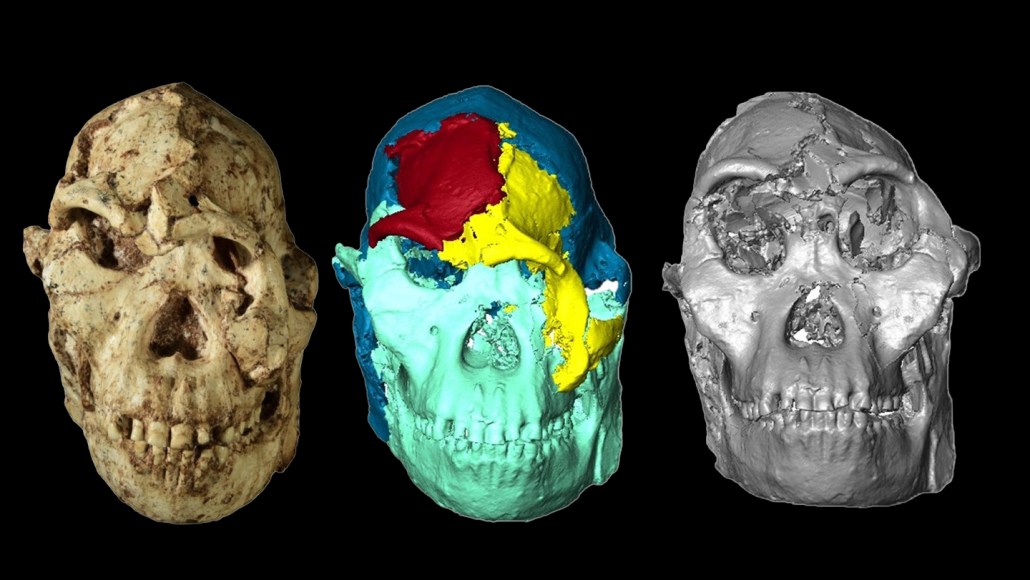
The skull was digitized using synchrotron X-ray imaging, producing a 3D dataset detailed enough to separate fragments virtually. Researchers then relied on supercomputers and semi-automated methods to isolate pieces and realign them into a coherent face, rather than attempting irreversible physical corrections. The CNRS description of the workflow emphasized that this was painstaking digital assembly, not a quick visualization step.
The resulting model reached 21 microns of resolution, and the CNRS said the reconstruction required more than five years of work. That resolution mattered because it supported fine-scale measurements across facial regions that had previously been too distorted to compare reliably. Once the face existed as a stable digital object, the fossil could finally be tested against other specimens with a consistent method.
The Numbers and Shape Tools Used to Compare Faces
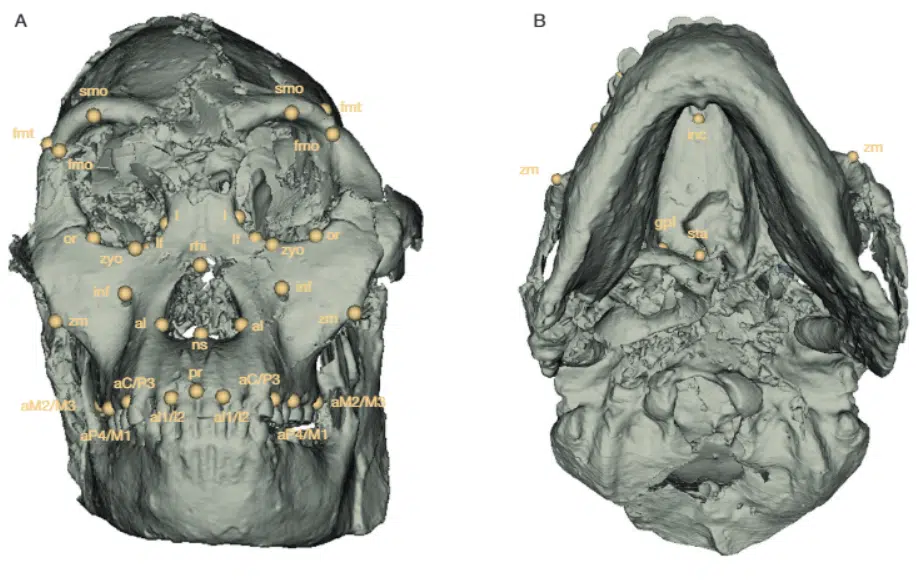
With the digital face reconstructed, the team measured it rather than simply describing it. They analyzed nine linear facial measurements and applied geometric morphometrics, a 3D shape-comparison approach that uses landmarks to evaluate overall form. The comparative set included living great apes and three other Australopithecus fossils, constrained by the basic reality that nearly complete fossil faces are rare.
Those three fossil comparisons included one younger specimen from South Africa and two Ethiopian specimens. The design of that comparison was deliberate: if geography neatly tracked facial similarity, a South African fossil might be expected to resemble another South African face most closely. Instead, the analysis tested that expectation using measured dimensions and computed shape relationships rather than visual impressions.

The reconstruction also allowed researchers to focus on a region that deformation had previously obscured. The team highlighted the orbital region, the bony structures around the eyes, as a key area where they saw evidence consistent with selective pressures. In the Wits University write-up, that point was tied to possible links between facial anatomy, visual capacity, and ecological behavior.
The Finding That Connected South Africa and East Africa
When the comparisons were run, the pattern did not follow geography. The Wits University release reported that the overall size of Little Foot’s face, the shape and dimensions of the eye sockets, and the general facial architecture more closely resembled the East African fossils than the younger South African comparative specimen.
It also carried a line that crystallized the surprise in plain language: “This pattern is unexpected, given the geographic origin of Little Foot and suggests a more dynamic evolutionary history than previously assumed,” says Beaudet.
First Appeared on
Source link

