Scientists Revive Brain Activity for the First Time After Seven Days in a Frozen Suspended State
Inside a laboratory in Germany, researchers cooled living brain tissue to temperatures colder than Antarctica’s harshest winter. The delicate samples were not preserved for storage or long-term archiving. Scientists instead wanted to test whether living neurons could survive a deep freeze that completely stopped their activity.
For seven days, the tiny brain slices remained at temperatures below −150°C. During that time, electrical signals inside the tissue stopped entirely. The microscopic connections that normally fire constantly inside living brains fell silent. At such temperatures, nearly all biological activity halts.
Then the researchers began slowly warming the tissue. The gradual process was designed to avoid sudden structural damage. Scientists monitored the samples closely during the transition out of the frozen state. The key question was whether the neurons would remain functional.
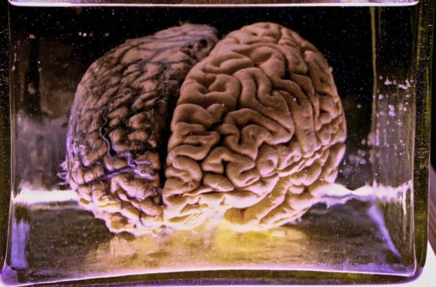
The work, published in a PNAS paper, was led by Alexander German of Friedrich-Alexander University Erlangen-Nuremberg. His team studies whether living brain tissue can survive controlled freezing. The experiment focused on slices of the hippocampus, a brain region closely tied to memory and learning. Detailed experimental results were later reported in Proceedings of the National Academy of Sciences.
Why Freezing Brain Cells Usually Fails
Freezing living cells is normally destructive. When temperatures drop, water inside cells forms ice crystals. Those crystals expand and puncture delicate cell membranes. The damage often leaves cells permanently unusable.
The brain is especially sensitive to this process. Neurons depend on fragile synapses that connect them into dense communication networks. Even small structural disruptions can block signals between cells. That vulnerability has long complicated attempts to freeze brain tissue safely.
Researchers have attempted similar experiments before. A well-known test in 2006 froze slices of rat hippocampus and later thawed them. The tissue survived structurally, but electrical signaling did not recover well. That outcome raised doubts about whether frozen brain circuits could ever fully function again.
The Glasslike Freezing Strategy
To avoid ice damage, the team used a technique known as vitrification. Instead of forming crystals, biological fluids solidify into a glasslike state. This state prevents the sharp structures that normally tear cells apart during freezing. Achieving vitrification requires carefully controlled cooling conditions.
The process relies on chemical mixtures called cryoprotectants. These substances reduce ice formation and stabilize cells during extreme cooling. Researchers treated slices of murine hippocampus with a carefully balanced cryoprotectant solution. The mixture aimed to protect neurons while limiting chemical toxicity.
After preparation, the samples were cooled rapidly to about −196 °C using liquid nitrogen. At that temperature, cellular processes essentially stop. The samples were then stored at −150 °C for seven days. During that period, the brain tissue remained in a vitrified state.
Microscopic inspection showed no visible ice crystal formation. That result suggested the cryoprotectant solution protected the tissue during freezing. Avoiding crystal formation was essential for preserving fragile neuronal structures. The next challenge was whether the tissue could recover.
The Signals That Returned
Researchers gradually warmed the samples to reverse the vitrified state. The careful warming process helped prevent structural stress inside the tissue. Once temperatures approached −10 °C, scientists began testing neuronal activity. The measurements focused on whether neurons could still communicate.
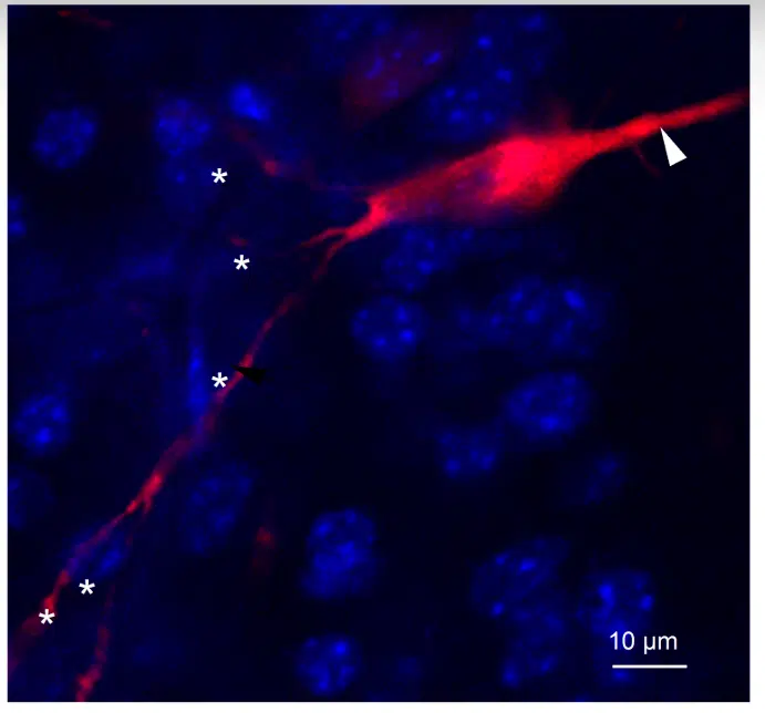
Electrophysiological tests revealed spontaneous synaptic events. These signals occur when neurons transmit messages across synapses. Their presence showed that communication between neurons had resumed. Electrical activity had returned after a full week of frozen suspension.
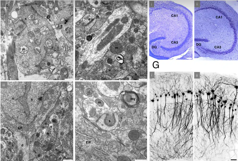
Microscopy also showed that many synaptic structures remained intact. The preserved connections allowed signals to move across neural circuits again. That recovery indicated that the tissue had survived the freezing process. Neural activity reappeared after the tissue warmed.
Why the Hippocampus Matters
The hippocampus was chosen because of its importance in forming memories. Its dense network of neurons makes it a demanding test for preservation techniques. If freezing damaged the network, electrical signals would not return. The restored activity therefore offered an important indicator.
The experiment did not directly test whether memories survived the freezing process. However, preserved synaptic activity suggests the physical wiring remained functional. Memory storage relies on patterns of synaptic connections. Maintaining those connections is essential for preserving neural information.
Future experiments will need to test more complex brain functions. Researchers may examine how long frozen tissue can remain viable. Larger sections of brain tissue could also be tested. Each step will help determine the limits of vitrified suspended states.
Toward Controlled Suspended Animation
Scientists have cryopreserved other organs before, including rat hearts and sections of liver tissue. The brain has remained far more difficult because of its fragile cellular networks. Even small disruptions can stop neurons from communicating. That complexity has slowed progress in brain preservation research.
The vitrification method developed at Friedrich-Alexander University Erlangen-Nuremberg avoids one of the biggest threats to cells. Preventing ice crystals protects neurons during extreme cooling. The approach allowed brain tissue to survive temperatures that stop all biological activity. The tissue later regained measurable electrical signals.
The experiment involved only small pieces of mouse brain tissue. Freezing entire organs or organisms would introduce additional challenges. Cooling larger structures evenly is much harder than freezing thin slices. Delivering cryoprotectants throughout a full brain would also be complex.
Still, the study showed that vitrification allowed mouse hippocampus tissue to remain frozen for seven days at −150°C and later recover electrical signaling after warming.
A. German, EY Akdaş, C. Flügel-Koch, E. Erterek, R. Frischknecht, A. Fejtova, J. Winkler, C. Alzheimer, & F. Zheng, Functional recovery of the adult murine hippocampus after cryopreservation by vitrification , Proc. Natl. Academic Sci. USA 123 (10) e2516848123, doi.org/10.1073/pnas.2516848123 (2026)
First Appeared on
Source link

